New High-Tech Microscope Boosts Cancer and Inflammation Research at UKSH Kiel
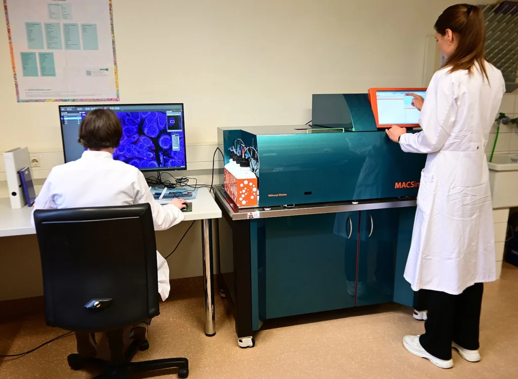
A major advancement in tissue analysis and new cutting-edge technology has arrived at the Section for Hematopathology at UKSH, Campus Kiel: The MACSimaTM system, a state-of-the-art immunofluorescence imaging platform, funded with €400,000 from the Kinderkrebsinitiative Buchholz/Holm-Seppensen (KKI) and the Medical Faculty of Kiel University, provides an unprecedented view of cellular interactions within tissue samples.
Traditional methods require separate stainings for different cell types, but the new system automates up to 60 stainings on a single sample. This allows researchers to study the spatial relationships between immune and tumor cells with far greater precision. Prof. Wolfram Klapper, head of the Hematopathology Section and project leader of P2 in the clinical research unit CATCH ALL, emphasizes its importance for cancer and inflammation research, as understanding these interactions is key to developing targeted therapies.
The technology will support major research initiatives, including CATCH ALL and the PMI Excellence Cluster, which focus on leukemia and chronic inflammation. As the first system of its kind in Kiel, the immunofluorescence imaging platform will serve as a shared resource, with standardized protocols enabling researchers to access this cutting-edge tool for translational medicine.
Read more about the microscope here (in German).